


Summary
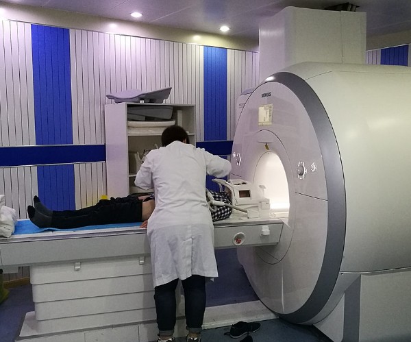
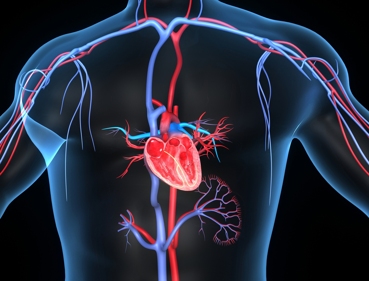
In this type of surgery, a pulse generator (called a pacemaker) is implanted in the chest to regulate the rhythm of the heart.
Preparation
Preparation for the procedure involves positioning the patient as required for the procedure, while the insertion site on the chest is carefully anaesthetised.
The patient is administered a sedative, which will make him or her feel relaxed and sleepy, while several monitors are connected to the patient’s chest and body.
The surgical care team will closely observe the monitors with data to monitor the patient’s heart rate and blood pressure during the procedure.
Driving Implant
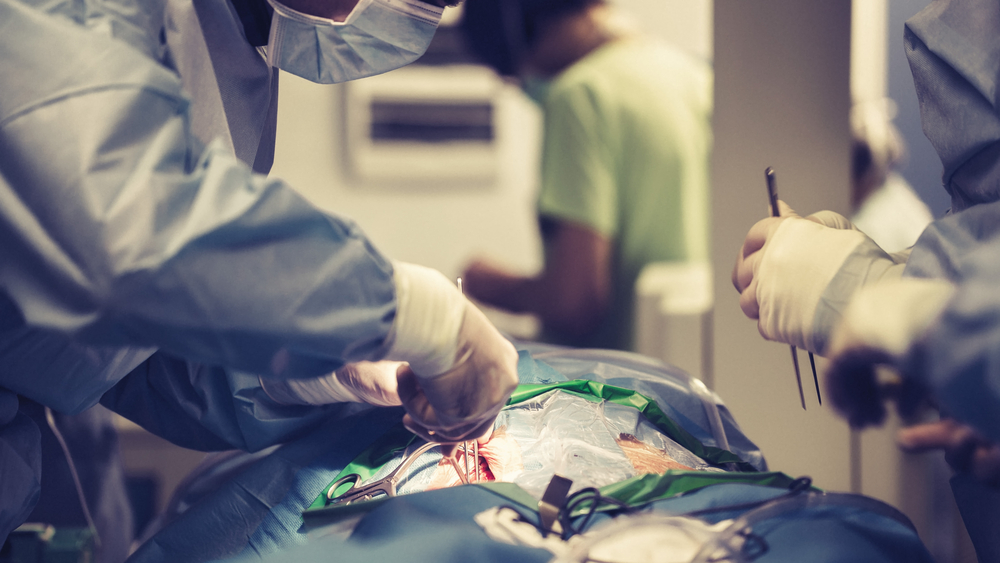
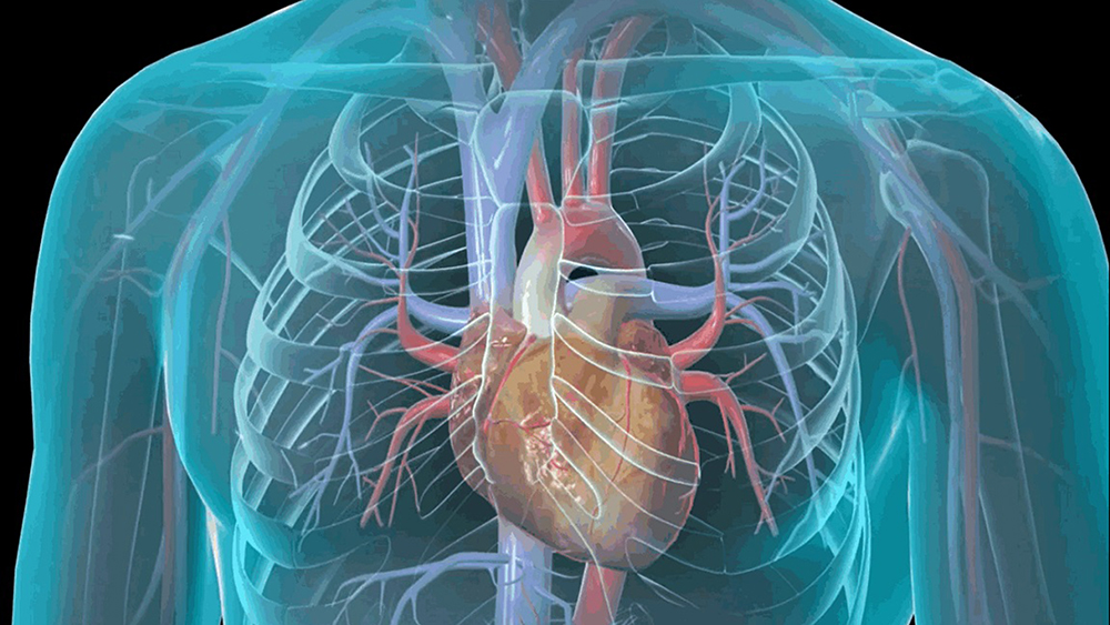
The doctor makes one or more small incisions to access a large vein in the chest in order to insert a lead, guiding it with careful precision into the heart with the help of a fluoroscope (a camera that creates a moving X-ray in real time).
Usually the lead is placed in the right ventricle of the heart. For some patients, two leads can be implanted (one in the right atrium and one in the right ventricle). This type of pacemaker is called a two-chamber pacemaker.
Testing the Guidance System
After the lead is in place, it is temporarily connected to a device that sends a series of impulses to the heart. This test is called ‘pacing’ and allows the doctor to ensure that the lead is working properly.
Pacemaker implantation
When the test has been performed, the test device is disconnected and the lead is connected to the pacemaker. The physician creates a small pocket under the skin of the chest and slides the pacemaker into this pocket. Once activated, the pacemaker will generate electrical impulses and send these signals through the leads and into the heart.
End of the procedure and subsequent treatment
When the implantation is complete, the doctor closes the incisions he had made and then adjusts the pacemaker settings with an external programming device to fine-tune the heart rhythm. The patient will remain in the hospital overnight so that the heart can be cautiously monitored closely.
Overview
In this procedure, an impulse generator (called apacemaker) is implanted in the chest to regulate therhythm of the heart.
Preparation
In preparation for the procedure, the patient ispositioned and the insertion site on the chest isanesthetized. The patient is given a sedative. This will make the patient feel relaxed and drowsy.Several monitors are attached to the patient’s chestand body. The surgical care team will closely watchthese monitors to track the patient’s heart rhythmand blood pressure during the procedure.
Implanting the Lead
The physician creates one or more small incisionsto access a large vein in the chest. The physicianinserts a lead into this vein and carefully guides itinto the heart with the help of a fluoroscope (acamera that creates a real-time moving x-ray).Typically, the lead is placed in the heart’s rightventricle. For some patients, two leads may beimplanted (one in the right atrium and one in theright ventricle). This type of pacemaker is called adual-chamber pacemaker.
Testing the Lead
After the lead is in position, the lead is temporarilyconnected to a device that sends a series ofimpulses to the heart. This test is called “pacing.” Itallows the physician to ensure the lead is workingproperly.
Implanting the Pacemaker
When the test is complete, the testing device isdisconnected and the lead is attached to thepacemaker. The physician creates a small pocketbeneath the skin of the chest and slips thepacemaker into this pocket. Once the pacemaker isactivated, it will generate electrical impulses andsend these signals through the leads and into theheart.
End of Procedure and Aftercare
When the implantation is complete, the physiciancloses the incisions. The physician may adjust thesettings of the pacemaker with an externalprogramming device to fine-tune the heart’s rhythm.The patient will remain in the hospital overnight sothe heart can be closely monitored.