Parkinson’s disease: gut microbiota may interfere with therapy

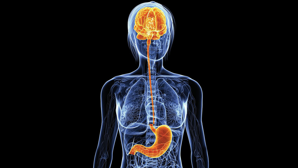
What does the gut microbiota have to do with a neurodegenerative condition such as Parkinson’s disease? Until yesterday, nothing, it was thought. Today, however, there seems to be a relationship, at least for those taking levodopa drugs to control tremors and muscle rigidity. That is, the vast majority of patients.

The role of gut bacteria

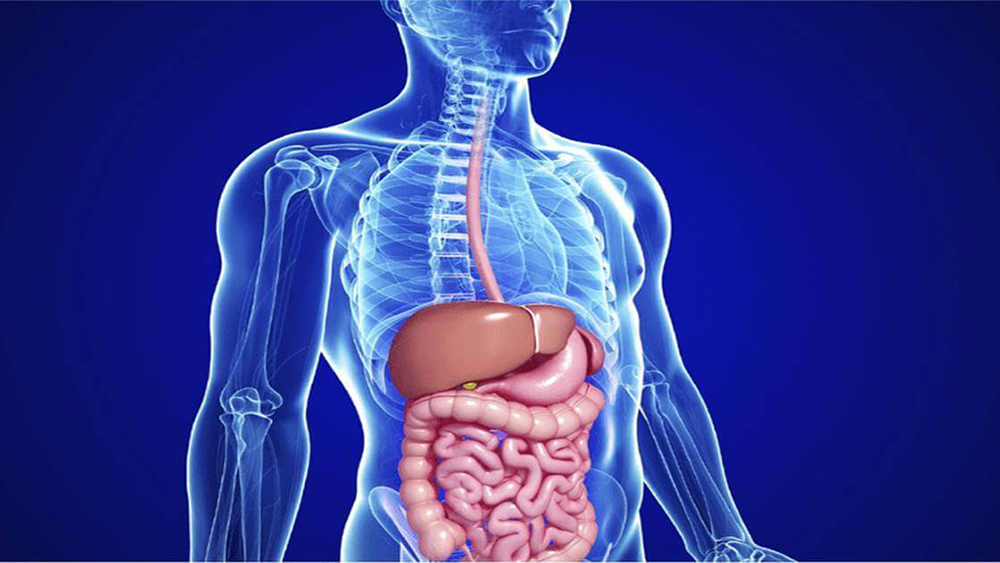
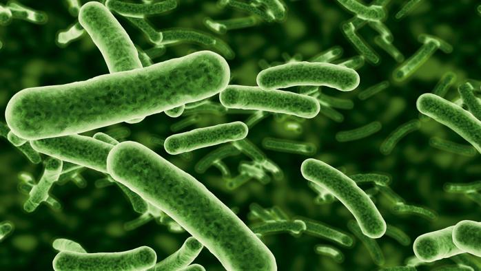
Intensive studies in recent years are teaching us that the gut microbiota serves as virtually everything, acting as an interface between us and the world and regulating countless enzymatic reactions and physiological functions. When healthy, everything is going well; when destabilized or depleted, the risk of seeing various ailments and diseases arise or worsen significantly increases.

In the case of
Parkinson’s,
the situation is somewhat different. Indeed, the characteristics and activity of patients’ gut bacteria do not create problems per se, but may interfere with the effectiveness of levodopa therapy: a dopamine precursor compound that must be constantly supplied from outside to replace dopamine no longer produced by the brain cells in the substantia nigra, degenerated due to the disease.

Levodopa is never administered on its own, but always in conjunction with a second substance called a “decarboxylase inhibitor,” which is essential so that levodopa is not rapidly converted into dopamine by the decarboxylases in the blood, but can reach the brain, where it must exert its action of modulating muscle tone.

It has been known for decades that, from the onset of the disease, different patients require different dosages of levodopa/inhibitors of decarboxylases to keep the symptoms of Parkinson’s disease under control and that, in all cases, the effective dosage gradually increases over time (due to a decrease in drug efficacy). Until now, however, no one could explain the reasons behind these phenomena.

New data on the effectiveness of therapy

A study recently published in Nature Communication suggests why this happens. In essence, the problem seems to be related to the ability of some intestinal bacteria (in particular, lactobacilli and enterococci, in which the constituent intestinal microbiota is rich) to transform levodopa into dopamine at the level of the small intestine, thus making it easily degraded soon after absorption and preventing sufficient amounts from reaching the brain.

Apparently, some Parkinson’s disease patients have a microbiota that is particularly active on this front, due to very high bacterial decarboxylase activity: this would make patients partially “resistant” to levodopa therapy, from which they might derive less than average benefits and gradually decrease over time.

Although it remains to be understood how to exploit these new data at the clinic-practice level, this is important information because it offers the possibility of improving and prolonging the efficacy of levodopa therapy by a different approach than those used so far, namely by modulating the composition of the gut microbiota and/or the activity of bacterial decarboxylases.

Source

Sebastiaan P et al. Gut bacterial tyrosine decarboxylases restrict levels of levodopa in the treatment of Parkinson’s disease. Nature Communications 2019;10:310. doi:10.1038/s41467-019-08294-y

Parkinson’s disease: Symptoms, causes and treatment

Parkinson’s disease is a chronic progressive disease; it was first described by James Parkinson in 1817. After dementias, it is the most common neurodegenerative disease.

EPIDEMIOLOGY

The condition is equally prevalent worldwide; the average age of onset is between
55 and 60 years of age
and affects
1-2% of the entire population over 65 years of age
; 5% of those affected are under 40 years of age.

In Italy there are about
220 thousand
people affected.

ETIOPATHOGENESIS

Various risk factors for Parkinson’s disease have been identified, including age, family history, male gender, environmental exposure to herbicides, pesticides, metals (manganese, iron), well water, residence rural, mental and physical trauma, emotional stresses.


A protective factor, on the other hand, is cigarette smoking.
.

The cause of the disease is unknown, but it is probably the result of an interaction between environmental toxins, genetic susceptibility, and senescence.
Mitochondrial dysfunction and oxidative stress
are now considered among the main mechanisms underlying the disease.

In a .small percentage
of the cases a genetic cause
. Major mutations include those affecting genes coding for
alpha-synuclein
and for
parkin
.

The
pathogenesis
is related to the
degeneration of most of the dopaminergic neurons in the black substance
(nerve structure located at the level of the midbrain). This results in the reduced production of
dopamine
, a key neurotransmitter in the
regulation of movement
. Symptoms of the disease become apparent
When more than 70 percent
of dopaminergic neurons were lost.

The main alterations involve the
black substance
which appears paler than normal; within it there is a reduction in the number of neurons and in surviving neurons inclusions of a substance called
alpha-synuclein:
are the so-called
Lewy bodies
, which are not specific to Parkinson’s disease, as they can also be found in
Lewy body dementia
and in
Alzheimer’s dementia.

SYMPTOMATOLOGY

The onset is sneaky
with tremor in one hand
, but also often with
joint pain
, depression of mood, easy fatigability.

The cardinal symptoms are
resting tremor
, the
slowness of movement
, also called bradykinesia,

and the
rigidity
. To these symptoms must be added postural instability. PD is a
asymmetric pathology
in that, especially in the early stages, it affects one half of the body more than the other .

Let us go on to illustrate the basic symptoms.

The resting tremor, present in about 70% of cases, has a frequency of 4-6 shocks per second and at onset affects only one hand and, in particular, the first three fingers, giving the impression of “count coins“; tremor is accentuated under conditions of emotional tension, fatigue, o When the subject feels observed, while it is absent during sleep.

The
rigidity
is characterized by an
increased muscle tone
with
constant resistance
to mobilization. There may be the so-called
cogwheel phenomenon
, in that the presence of resistance to passive mobilization is alternated with
sudden failures
, brings to mind the
clicks of a gear
.

The
slowness of movement
(bradykinesia) is the third cardinal sign of the disease, also accompanied by reduced motility. During the march, the subject with PD gives the impression of having the
upper limbs attached
to the body
without the characteristic
pendular movements
.

Also observed are
difficulties such as using a knife or fork
, buttoning up or
unbuttoning
, shave. The writing becomes trembling and uncertain and you shrinks (microgra phy), the facial expressions is reduced (amimia) (Figure 8). The subject, over time, assumes a prone posture with
Head flexed forward and knees and elbows flexed
.


Postural instability
is the difficulty in maintaining upright station in response to external thrusts; it can be the cause of disastrous falls and is present in about 40% of cases.

NON-MOTOR SYMPTOMS

Autonomic nervous system involvement is characterized by the presence of
orthostatic hypotension, constipation, sialorrhea, seborrhea, increased sweating
At the level of the head and neck. Urinary disorders may be present such as urinary urgency
and increased
urinary frequencyI
.

The
cognitive impairment
, usually moderate, is present in up to 60 percent of cases; there may be impairment of attention, concentration and memory and slowness in performing executive tasks. Unfortunately in some cases until the
in 20%
may be present a
frank dementia
subcortical type

The
depression of mood tone
is often present but under-diagnosed and under-treated. I sleep-wake rhythm disorders are very common being able to affect up to 90% of people with PD. They can consist of
excessive daytime sleepiness
or in
difficulty in initiating or maintaining sleep
, or in
poor sleep quality with frequent awakenings
and reductions in stages III and IV sleep and REM sleep.

DIAGNOSIS

It is mainly based on the recognition of the three main signs
, resting tremor, rigidity and bradykinesia with unilateral onset, and the response to L-DOPA.

CT or MRI scans of the brain are normal or at most can demonstrate the presence of some degree of brain atrophy, which is otherwise highly variable.

The following can be used to assess presynaptic dopaminergic pathways
the SPET-DaT SCAN, which s
erve to show alteration of dopaminergic pathways, but is not always superior to clinical diagnosis.

To assess the degree of disability, the use of Rating Scales is useful. The most widely used are the UPDRS and the Hoehn and Yahr Scale.

THERAPY

Initially there is a good drug response; later, as the disease worsens, the response to drugs worsens and fluctuations appear especially in the area of motor symptoms. These include the on-off phenomenon, sudden lack of response to medication with no relation to the timing of intake; wearing-off, predictable reappearance of parkinsonian symptoms after a number of years due to the shorter duration of response to medication; and freezing of gait or simply freezing, sudden motor blockage that occurs at the onset of walking, or in crossing narrow passages, or in changes of walking direction; in the latter case, the subject reports having his or her feet as if glued to the floor.

Some drugs used in Parkinson’s disease stimulate creativity

In individuals with Parkinson’s disease, the use of dopaminergic drugs that can stimulate receptors for dopamine, a deficient neurotransmitter in this disease, can result in side effects characterized by poor impulse control: these include pathological gambling, hypersexuality, and compulsive shopping. However, the same drug treatment has stimulated increased creativity and the acquisition of new artistic skills in some individuals with Parkinson’s disease.

While poor impulse control, such as especially pathological gambling, is well recognized in the literature, creativity remains underestimated, probably because it is an often-appreciated event and as such not likely to create distress, either in patients or their families. In a recent study, researchers evaluated about 20 subjects with Parkinson’s disease who exhibited greater artistic creativity after dopaminergic treatment than before. Among the artistic activities found in first place is painting, but also the development of poetic skills(Schrag and Trimble 2001; Walker et al, 2006).

There is no doubt that a stimulating family environment could bring out creative aspects more frequently during Parkinson’s disease treated with dopaminergic drugs. But a not insignificant aspect is that the same patients in whom there is the appearance of artistic activities also report a state of well-being and a loss of awareness of the disease and even of disease-related physical limitations(Chatterjee et al, 2006).

Source: Garcia-Ruiz PJ, Martinez Castrillo JC, Desojo LV. Creativity related to dopaminergic treatment: A multicenter study. Parkinsonism Relat Disord. 2019 Feb 22. pii: S1353-8020(19)30056-2.

Bibliography

Chatterjee A, Hamilton RH, Amorapanth PX: Art produced by a patient with Parkinson’s disease. Behav Neurol 2006;17:105-108

Schrag A, Trimble M: Poetic talent unmasked by treatment of Parkinson’s disease. Mov Disord 2001;16:1175-1176

Walker RH, Warwick R, Cercy SP: Augmentation of artistic productivity in Parkinson’s disease. Mov Disord 2006;21:285-286.

Is it possible to stop the progressive worsening of Parkinson’s disease?

The
Parkinson’s disease
is a degenerative disease of the central nervous system, first described in 1817 by an English physician, James Parkinson. It is characterized by three main symptoms: slowness of movement, rigidity and tremor. The causes are related to degeneration of certain structures in the central nervous system, where dopamine, the main neurotransmitter essential for controlling bodily movements, is produced. Treatment of the disease still relies on drugs that can either supply the deficient dopamine through its precursor, L-DOPA, or stimulate the cells on which this transmitter acts, the dopaminergic cells. Unfortunately, due to the processes of neuro-degeneration, inherent in Parkinson’s disease, these drugs gradually lose their effectiveness over time.

A pioneering program involving an experimental and innovative drug treatment to be applied directly to the brains of people with the disease was published in the latest issue of the Journal of Parkinson’s Disease in February 2019.

The hope: to restore cells damaged in the course of the disease.

The study is based on providing Parkinson’s disease-compromised brains with increased levels of a naturally occurring growth factor, glial cell line-derived neurotrophic factor (GDNF), which has been shown to be able to regenerate the brain cells impaired dopaminergic pathways in individuals with this condition.

Six patients took part in the initial pilot study that mainly evaluated the safety of the treatment approach. Another 35 people then participated in the actual study, which was carried out in a double-blind manner for the duration of nine months: half of the randomly selected subjects received monthly infusions of GDNF, while the other half were treated with placebo infusions.

A specially designed delivery system using robot-assisted neurosurgery was implanted in each subject to carry out the monthly infusions. This delivery system made it possible to deliver high-dose infusions of GDNF every four weeks directly to disease-affected brain areas with pinpoint accuracy through a transcutaneous port mounted on the skull behind the ear. The high compliance rate (99.1%) in participants recruited throughout the United Kingdom demonstrated that the drug delivery system, by repeated cerebral infusion, is feasible and tolerable.

After 18 months of therapy, all of the patients who had received GDNF showed improvement in disease-affected brain areas and related symptoms with a moderate to major rating from the researchers compared with the initial condition. This improvement was also observed in those subjects who were initially placed in the placebo group and then switched to GDNF treatment. GDNF administration proved to be safe throughout the study period.

The study’s principal investigator, Dr. Alan L. Whone of Bristol Medical School at the University of Bristol, UK, stated that: “ In GDNF-treated subjects The improvement in disease-affected areas went beyond what has ever been seen before” and then added that: “High doses of GDNF are able to awaken and restore dopaminergic brain cells, which are progressively impaired in the course of Parkinson’s disease.”.

Even in light of the neurodegenerative process underlying Parkinson’s disease, the drugs currently in use are likely to gradually lose their effectiveness. Therefore, there is no doubt that this study, if confirmed by subsequent evaluations, represents a decisive breakthrough in the treatment of Parkinson’s disease.

Source: Alan L. Whone et al:“Extended Treatment with Glial Cell Line-Derived Neurotrophic Factor in Parkinson’s Disease” published online in the Journal of Parkinson’s Disease, in advance of Volume 9, Issue 2 (April 2019) by IOS Press

Visual dysfunction, a misrecognized symptom of Parkinson’s disease

“The idea that visual symptoms may be associated with Parkinson’s disease is not new, but this is the first time it has been reported at the population level,” he told Medscape Medical News Ali Hamedani of the University of Pennsylvania in Philadelphia, lead author of a study published in European Journal of Neurology.
Analyzing data obtained through a large survey, U.S. researchers found that visual dysfunction is significantly more common in individuals with Parkinson’s disease than in the general adult population. However, this is a generally overlooked and untreated factor, even though it worsens the already compromised quality of life of these patients.
The survey was conducted on more than 150,000 individuals and calculated that people diagnosed with Parkinson’s disease were more than twice as likely to suffer from vision impairment in both near and far vision as the general population.
Vision problems had been repeatedly reported by the patients themselves, their caregivers and attending physicians. So, the suspicion was evidently already there, but the new study provides a confirmation supported by a large-scale survey, and the large number of data allows a statistical correlation to be documented that otherwise would not have been so stringent.
With colleague Allison Willis, Hamedani analyzed data of adults aged 50 years and older provided by the Survey of Health, Aging and Retirement in Europe. (SHARE), a multinational population health survey of people living in one of 27 European countries and Israel.
Although the risk of impaired distance vision was 2.55 higher (and near vision 2.07), people with Parkinson’s reported this difficulty to an ophthalmologist less often, so that the condition remained untreated in many cases.
The study authors report that there have been some suggestions of a subtle change in color vision in the years before Parkinson’s diagnosis, but it is unlikely that patients themselves will notice. “It could probably be found out if screening was done, but it is too early for any recommendations on that,” Hamedani said. Instead, the researcher suggested that physicians examine visual function in already diagnosed patients, although the nature and significance of these symptoms still appear unclear: we do not know whether the mechanism is the result of Parkinson’s-associated changes directly in the eyes or at the brain level.
Source:
Hamedani AG, Willis AW. Self-reported visual dysfunction in Parkinson disease: the Survey of Health, Ageing and Retirement in Europe. European Journal of Neurology 2019, 0: 1-6.

Parkinson’s disease, how Facebook founder proposes to treat it

Considering using an intercranial device (Wand) , wireless, capable of recording, stimulating and modifying a monkey’s movements in real time, which once implanted in a human brain could help treat diseases such as epilepsy and Parkinson’s disease.
The idea ties in with the largest investment of the Chan Zuckerberg Initiative, a project created and supported by the Facebook founder and his wife Priscilla Chan, with the important goal of “curing all existing diseases.”

The project

The capital earmarked for the project amounts to the considerable sum of $5 billion, cash flow from a sale of as many as 29 million Facebook shares.

The small Wand device , among the first co-funded projects ,whose tests have already been carried out on monkeys, was the subject of a scientific study later published in the scientific Journal Nature Biomedical Engineering.

The device

UC Berkeley in conjunction with startup Cortera has created Wand, which consists of a small brain pacemaker that can transmit signal from 128 different points in the brain with the help of electrodes and deliver electrical discharges that can influence behavior.

The test conducted on monkeys saw the animals intent on moving a cursor on a screen using a joystick, an action they could perform because of previous training.

Wand modified the animals’ movement by sending an electrical signal into the brain,

The ‘possible use

It is hoped that Wand may be a valuable contribution to treating a range of movement disorders, such as spinal cord injury and epilepsy, due specifically to the ability to anticipate motor action, with the ability to stop it.

Similar projects, such as Elon Musk‘s Neurali, had been started before such as the famous Building 8, also by Facebook, but today this procedure can be conducted , as a relevant novelty, without the use of wires, avoiding that physical link between machine and brain.

CULTURA E SALUTE
 
AGGIORNAMENTI
 
PERCORSI
 

your advertising
exclusively ON
MY SPECIAL DOCTOR

complete the form and you will be contacted by one of our managers