ARTICOLI CORRELATI

Why do I get cramps when I run?

A muscle cramp can come suddenly , such as during a run , and it does not announce itself with any kind of sign. At most you may realize it is coming very late , when already the muscle is contracting or overlapping.

Application of Stents (Coronary Angioplasty)

A coronary stent is a medical "device," that is, an instrument used in Coronary Angioplasty to clear the way for possible blockages in the coronary arteries by following invasive techniques having the goal of restoring regular blood circulation within the heart by ensuring its proper function.

Hypertrophic cardiomyopathy

Hypertrophic cardiomyopathy is a condition in which the heart muscle thickens, becoming hypertrophic, in the absence of dilation of the ventricles. Hypertrophic cardiomyopathy affects men and women equally. It is

Application of Pace-Maker

Each pace-maker consists of two basic parts: a pulse generator and one or two electrodes that are run from the generator to the heart muscle.

Diving, the basic rules

The human body, while moving through water, swimming or diving, knows that it poses a constant danger since it cannot survive under this element except with reserves of air. Never dive alone.

Working professions in mitral valve disease

It is well known that risk factors for heart disease include habits such as excessive alcohol use, such as smoking, a high-protein and high-fat diet, and also genetic factors, while less consideration is often given to factors of occupational origin.

CT Computed Axial Tomography

tac

Computed tomography, abbreviated as CT, still often commonly referred to as CAT (computerized axial tomography) despite the evolution of the method, is an imaging investigation that provides very detailed three-dimensional structural information of the internal organs of the human body, making use of ionizing radiation (X-rays).

Description

To perform the CT scan, it is, first of all, necessary to remove all metal or other objects (earrings, rings, bracelets, chains, glasses, etc.), undress and put on the gown provided by the health care personnel.

Then, one should lie down on the couch of the CT apparatus and get into the position indicated by the doctors, which should be held stably throughout the evaluation, following precisely the doctors’ requests regarding breathing. When the correct position has been assumed, the crib begins to slide, going through a structure similar to a large doughnut with a hole in it, stopping in places at the areas to be analyzed.

At this stage, the CT scan circle administers ionizing radiation while special sensors acquire signals from the body tissues examined; in doing so, the circle rotates on itself to acquire information in all directions in space.

The collected signals are transduced into electrical pulses and sent to a computer that integrates and processes them, turning them into three-dimensional images, displayed on the monitor, which can be further processed to optimize their resolution and photographed to fix the most interesting aspects for clinical purposes.

The duration of the CT scan varies according to the extent and complexity of the area of the body to be examined (encephalon, chest, abdomen, limbs, etc.), as well as the type of instrument used (newer or older) and the patient’s ability to maintain the required position. On average, the total duration of the evaluation is about 30 minutes. The examination is completely painless.

In some cases, to improve the diagnostic ability of the CT scan, a contrast agent is to be used, which, depending on the case and the purpose of the investigation, will have to be taken by mouth (liquid) or injected into a vein or inserted into the rectum, a few minutes before lying down on the instrument bed.

When needed

Now widely used in clinical practice, CT scanning is considered a Level 2 evaluation, to be used when other simpler, cheaper, and harmless techniques do not allow to specify the diagnosis with a sufficient degree of accuracy and in planning interventional procedures.

One of the main reasons for limiting the use of CT scans relates to the fact that the proportion of ionizing radiation administered during the examination, while not harmful when considering a single evaluation, is not negligible (amounting to about 30 times that used in performing X-rays), and the accumulation of the effects of X-rays administered in numerous subsequent CT scans, even years apart, could damage the body’s tissues and promote the development of fibrosis or tumors.

The main indications for performing CT include:

  • Diagnosis of muscle and bone diseases, such as sarcomas, bone tumors, and complex fractures;
  • Clarification of the location (and relationship to neighboring tissues/organs) of a tumor, site of infection, or blood clot (thrombus);
  • Planning for an invasive procedure such as a delicate surgery or biopsy;
  • Accurate identification of the area where a tumor is present to be treated with radiation therapy;
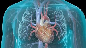
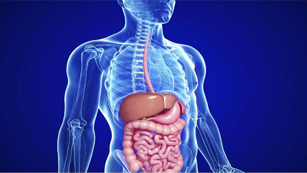
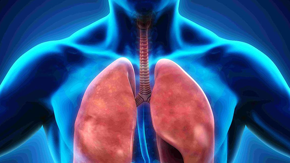
  • Detection and monitoring of tumors, heart disease (coronary angiography with CT), lung nodules, and liver masses;
  • Evaluation of the effectiveness of specific therapies, particularly cancer therapies;
  • Accurate detection and localization of injuries and bleeding to internal organs.

Special Warnings

By itself, CT scan has no particular contraindications. However, in view of the use of ionizing radiation, it is important for women of childbearing age to be certain that they are not pregnant at the time the examination is performed. Conversely, although the radiation dose used is not likely to harm the fetus, if possible, it is preferable to postpone the evaluation or opt for an alternative diagnostic method (ultrasound or MRI).

The contrast medium used for CT scanning is generally harmless, but some people may develop hypersensitivity reactions (with onset of itching and skin rash) or true allergies: if you have had such experiences in previous examinations with contrast medium (even other than CT) or you are intolerant or allergic to particular drugs, it is important to inform your health care provider already while scheduling your CT appointment.

SPECIALISTI IN EVIDENZA

  • Profile picture of Dr. Giuseppe Lepore
    active 4 years, 1 month ago

    Dermatologists, Basic Doctors, General Surgeons

    • 19 Via Manzoni Alessandro - Garbagnate Milanese
    phone
  • Profile picture of Studio Medico San Pellegrino
    active 3 years, 10 months ago

    Sports Doctors, Basic Doctors

    Spread the word!

    Recommend this special

    doctor to a friend

    message phone house
    visit
    video
    consulting
    WhatsApp conferences
    and webinars
  • Profile picture of Dott.ssa Floriana Di Martino
    active 4 years, 1 month ago

    Aesthetic Doctors, Basic Doctors

    • Piazza della Rinascita 13 - Pescara
    phone
  • Profile picture of Dott. Flavio Della Croce
    active 4 years, 1 month ago

    Psychotherapists, Basic Doctors

    • Via Sacconi 1 - Borgonovo Val Tidone
    phone
  • Profile picture of Dr. Domenico D'Angelo
    active 4 years, 1 month ago

    Otolaryngologists, Basic Doctors, Competent Doctor

    • Via Pola 5 - San Prisco
    phone

PATOLOGIE CORRELATE

Coronaropathy

The term coronary artery disease refers to chronic “distress” of the coronary arteries, the arteries that supply blood to the heart muscle, in most cases brought about by atherosclerotic pathology

Aortic valvulopathy

When the aortic valve does not properly perform its function of pumping blood into the heart, an aortic disease condition occurs that also involves the left ventricle. Aortic valve disease

Jugular stenosis

Jugular stenosis corresponds to a narrowing of the lumen of the jugular veins, which are the main blood vessels that collect oxygen-poor blood from the brain and return it to

Mitral valve repair

Repair of an insufficient/deteriorated mitral valve can be done: Removing the malfunctioning segments of the valve leaflets; implanting “synthetic cords” (which replace the broken or “stretched” natural ones of the

Heart failure

Heart failure corresponds to a condition in which the heart is unable to receive and/or pump blood with sufficient force to the lungs and the rest of the body, due

Ventricular hypertrophy

Ventricular hypertrophy is a condition characterized by thickening and a loss of elasticity of the walls of the left ventricle (i.e., the left lower chamber of the heart), which thus

Hypertension

According to the ESH-ESC guidelines, hypertension is defined by the finding of systolic (maximum) blood pressure values ≥ 140 mmHg and/or diastolic (minimum) blood pressure values ≤ 90 mmHg. The

Cardiac Echodoppler

The echocardiogram, also known as echocardiography, is a diagnostic imaging technique based on the use of ultrasound, which is quick and easy to perform, harmless, painless, and low-cost, and because

Venous insufficiency

Venous insufficiency and associated vascular disorders, such as varicose veins and telangiectasias (superficial capillaries), mainly affect the venous vessels of the legs and arms and are mainly related to the

Deep vein thrombosis (DVT)

Deep vein thrombosis (DVT) occurs when a clot (thrombus) forms within a vein and completely or partially occludes it, blocking or restricting blood flow in that vessel. In principle, any
CULTURA E SALUTE
 
AGGIORNAMENTI
 
PERCORSI
 

your advertising
exclusively ON
MY SPECIAL DOCTOR

complete the form and you will be contacted by one of our managers