ARTICOLI CORRELATI

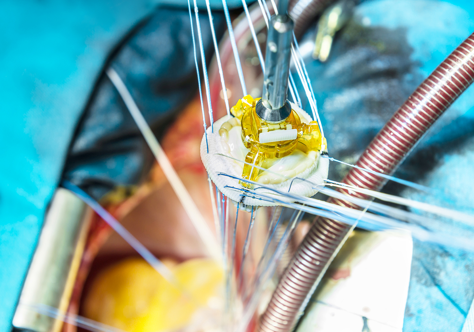
Aortic valve replacement

When the condition of the aortic valve is severe and the insufficiency is exposing the patient to heart failure, as well as the possibility of developing other, serious, life-threatening conditions, aortic valve replacement is the way to go.

ERECTILE DYSFUNCTION, WHAT TO KNOW AND WHAT TO DO

The mechanism that determines erection in the male arises from sexual arousal, which in turn is the result of the participation of emotions, the brain, hormonal flows, heart and vessels, muscles and nerves. Sometimes even one of these factors can cause erectile dysfunction, which can worsen when a state of emotional malaise and stress or even a persistent state of anxiety is added to it.

Heart failure: disorders of the heart

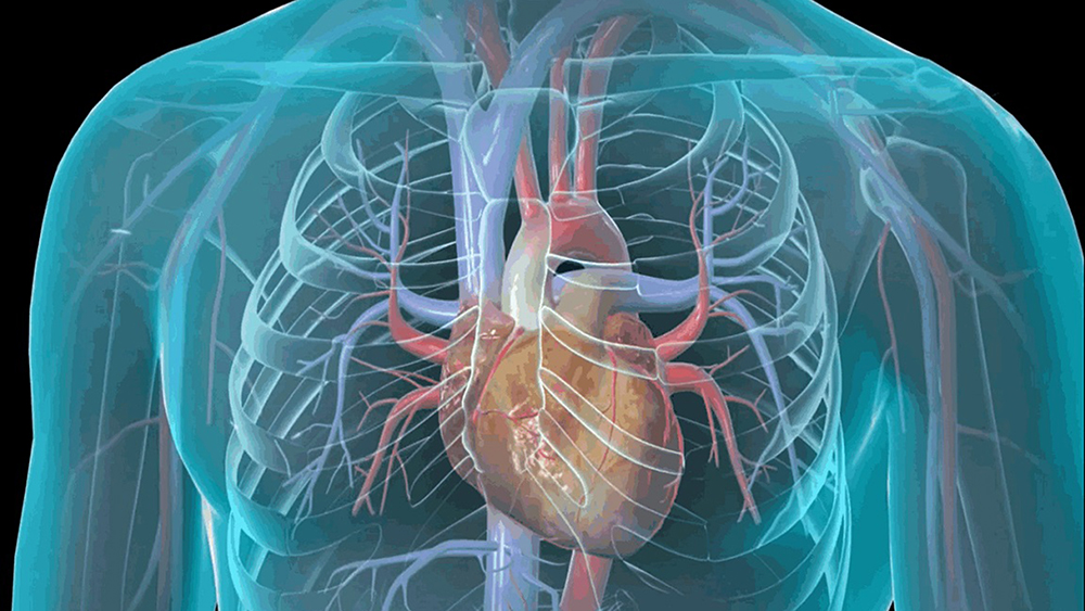
Heart failure refers to a dysfunction as a result of which the heart can no longer meet the needs of the body, causing reduced blood flow and accumulation (congestion) of

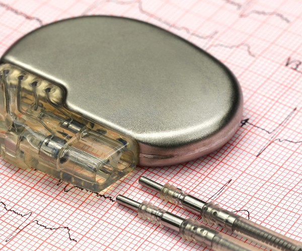
Application of Pace-Maker

Each pace-maker consists of two basic parts: a pulse generator and one or two electrodes that are run from the generator to the heart muscle.

Too much Caffeine?

Taken in small doses it improves concentration, while if taken in excessive doses it causes heart rate, raises blood pressure levels and induces a state of nervousness.

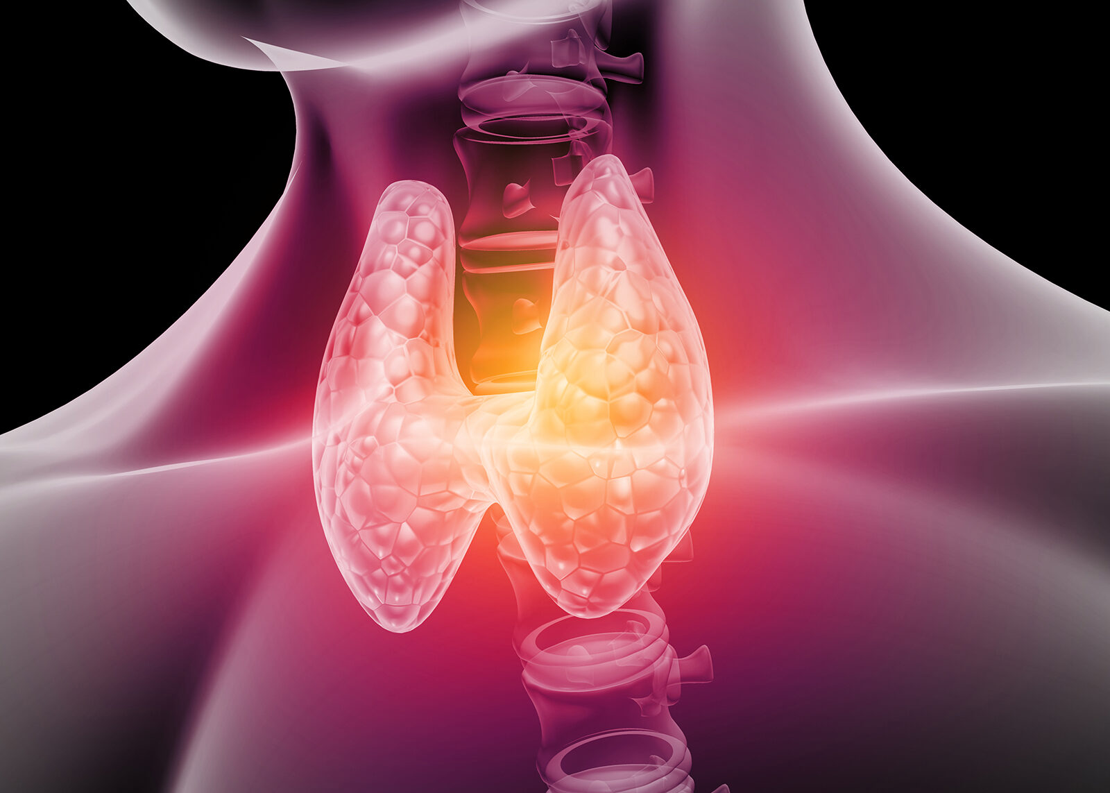
Hashimoto’s thyroid

When the immune system launches an attack on the thyroid gland, the small gland located under the Adam's apple, which produces hormones that regulate so many functions of the human body.

Aortic valve repair

When aortic valvulopathy presents significant quality-of-life problems for the patient but also obvious risks of serious consequences, cardiac surgery may be necessary.

Echocardiogram

ecocardiogramma

The echocardiogram, also known as echocardiography, is a diagnostic imaging technique based on the use of ultrasound, which is quick and easy to perform, harmless, painless, and low-cost, and because of these favorable features has become very popular in the last 20 years in cardiology to study the heart, the blood vessels surrounding it, and the heart valves.

Description

An echocardiogram is for all intents and purposes an ultrasound scan performed at the level of the heart that can be conducted transthoracic or transesophageal and allows visualization of the heart, the blood vessels surrounding it, and the heart valves during their normal functioning, at rest, after stress testing, or after taking a drug, providing two- or three-dimensional images.

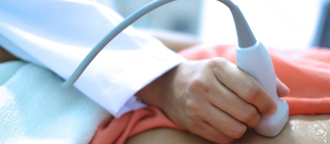
The transthoracic mode is the simplest and is performed similarly to a common external ultrasound, scanning the chest with a sensor/transducer after applying a special gel to the clean, dry skin that optimizes ultrasound transmission to and from the structures to be examined.

The sensor/transducer sends diagnostic ultrasounds, which pass through the body tissues for several centimeters and are partially “reflected” by them back to the sensor/transducer, differentially according to the characteristics of the tissue itself. The signals received and transduced by the sensor into electrical pulses are sent to the ultrasound scanner, which translates them into images (black and white or color), displayed on the monitor in real time.

This makes it possible not only to visualize all the structures in the area scanned by the ultrasound scanner, but also to assess the contraction of the heart, the opening/closing of the valves, and the flow of blood as they function.

The transesophageal modality is a bit more complex and involves mild preliminary sedation and the insertion into the esophagus of a small tube with the sensor/transducer on the end, which is placed at the level of the heart. From here, having to pass through minimal layers of well-hydrated tissue, the ultrasound arrives and is reflected off the structures to be examined more efficiently and with less interference, providing more detailed and better defined images.

Regardless of the modality used, during the course of the examination, the physician takes a number of snapshots of the images on the monitor that he or she considers to be of particular relevance for diagnostic purposes. Examination by transthoracic modality takes about 15-20 minutes on average, while that conducted by transesophageal modality is somewhat more challenging, partly because of the time required for initial sedation and final recovery. In both cases, the report is usually delivered immediately after the evaluation is finished.

When needed

The echocardiogram is performed to assess the size, structure, and dynamic function of the heart, heart valves, and related vessels in the presence of clinical symptoms suggestive of the presence of heart disease and/or after other first-level cardiologic examinations (e.g., electrocardiogram, ECG) have resulted in findings worthy of further investigation. In addition, the test is used to monitor already diagnosed heart disease and before/after surgery. The main conditions that lead to the indication of performing the echocardiogram include:

  • heart failure;
  • alterations in heart rhythm;
  • dilated cardiomyopathy;
  • coronary artery disease;
  • altered anatomy and malfunction of heart valves;
  • congenital heart defects;
  • outcomes of cardiac surgery.

Special Warnings

Performing the echocardiogram involves no special preparation. If you are already on medications to control known heart disease, your physician will indicate when to take them in the hours before the exam to optimize the outcome of the evaluation.

If the examination is conducted transesophageally, a mild sedation, which is generally harmless, is expected to be administered in advance: in this case, any allergies/intolerances to certain medications and a complete list of all medications, nutritional supplements and phytotherapeutic/alternative remedies that are being taken should be communicated to the physician in advance in order to avoid possible unfavorable interactions.

However, the list of all substances taken (including alcohol and beverages containing caffeine or other stimulants) should always be reported to the physician so that he or she can better interpret the outcome of the evaluation and more easily trace the possible cause of the abnormal cardiac symptoms and signs found.

Echocardiogram can be performed at any age, including pediatric age.

SPECIALISTI IN EVIDENZA

  • Profile picture of Dott. Cristiano Crisafulli
    active 3 years, 11 months ago

    Diabetologist, Basic Doctors, Internist

    • Via Vittorio Emanuele II 181 - Acireale
    phone
  • Profile picture of Dr. Giovanni Arrichiello
    active 3 years, 11 months ago

    Gynecologists, Basic Doctors

    • Corso Regina Margherita 260 - Torino
    phone
  • Profile picture of Dott. Sandro Morganti
    active 3 years, 11 months ago

    Orthopedists, Basic Doctors, Osteopaths

    • Via Foligno 6 - Roma
    phone
  • Profile picture of Dott.ssa Floriana Di Martino
    active 3 years, 11 months ago

    Aesthetic Doctors, Basic Doctors

    • Piazza della Rinascita 13 - Pescara
    phone
  • Profile picture of Dr. Giuseppe Lepore
    active 3 years, 11 months ago

    Dermatologists, Basic Doctors, General Surgeons

    • 19 Via Manzoni Alessandro - Garbagnate Milanese
    phone

PATOLOGIE CORRELATE

Atherosclerosis

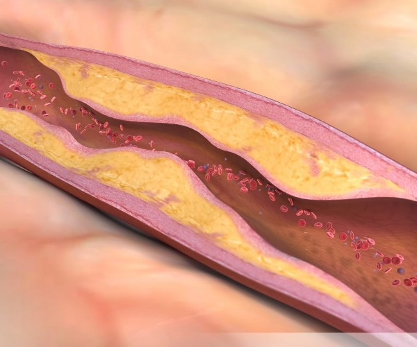
Atherosclerosis is a pathological process that leads to the progressive formation of localized deposits composed mainly of fat and cholesterol and platelets (atheromas or atherosclerotic plaques) within the walls of

Shock, medical emergency

It may happen that the circulation of blood through the body undergoes a sudden change giving rise to a condition of physiological shock, with decreased flow and crisis on the

Venous insufficiency

Venous insufficiency and associated vascular disorders, such as varicose veins and telangiectasias (superficial capillaries), mainly affect the venous vessels of the legs and arms and are mainly related to the

Hypertension

According to the ESH-ESC guidelines, hypertension is defined by the finding of systolic (maximum) blood pressure values ≥ 140 mmHg and/or diastolic (minimum) blood pressure values ≤ 90 mmHg. The

Cardiac arrest

Cardiac arrest corresponds to the interruption of the heart’s contraction and pumping action, resulting from a sudden and drastic alteration in its electrical activity (ventricular fibrillation). Cardiac arrest is an

Blow to the heart

A “heart murmur” is not in itself a pathology , but merely a signal that the blood inside the heart is flowing too fast or in a “disordered” manner, producing

Mitral valve repair

Repair of an insufficient/deteriorated mitral valve can be done: Removing the malfunctioning segments of the valve leaflets; implanting “synthetic cords” (which replace the broken or “stretched” natural ones of the

Angina pectoris

Referred to by the Latin name for its main manifestation, a weight-like or vice-like tightness in the chest, angina pectoris is not a disease per se, but the consequence of

Cellulite

Cellulite is an inflammatory-based alteration of the fat-rich subcutaneous tissue(panniculus adiposus), triggered by fluid stagnation due to poor venous and lymphatic circulation (lymph is a fluid that flows in channels

Ventricular hypertrophy

Ventricular hypertrophy is a condition characterized by thickening and a loss of elasticity of the walls of the left ventricle (i.e., the left lower chamber of the heart), which thus
CULTURA E SALUTE
 
AGGIORNAMENTI
 
PERCORSI
 

your advertising
exclusively ON
MY SPECIAL DOCTOR

complete the form and you will be contacted by one of our managers