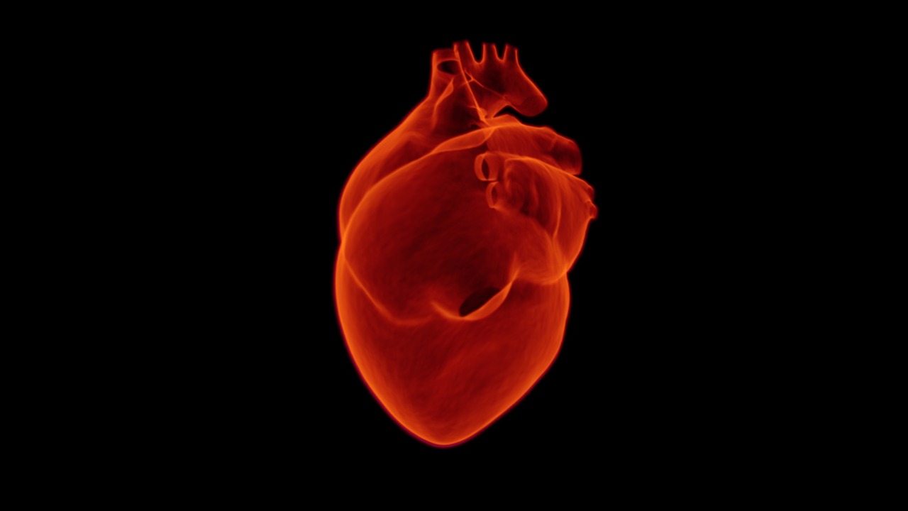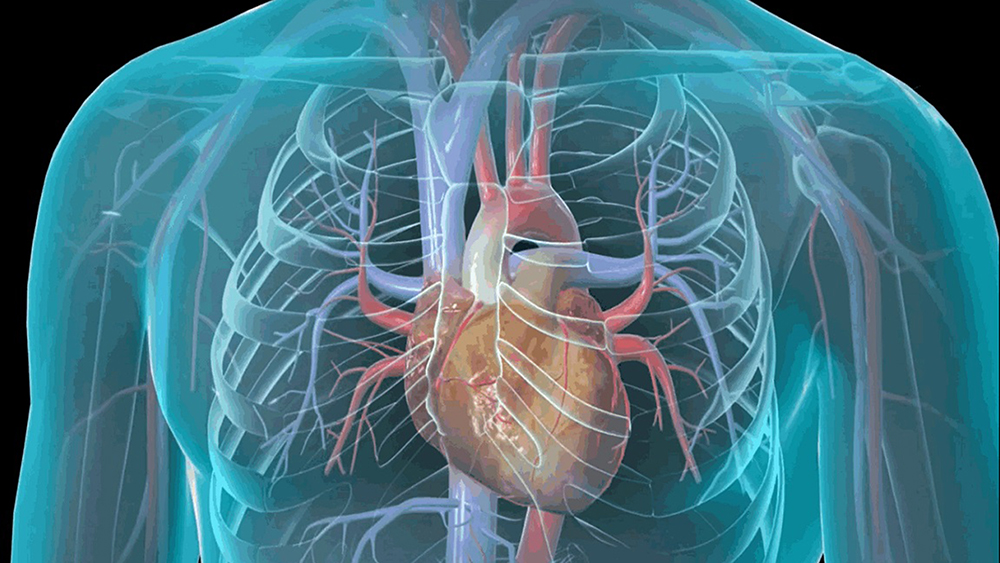


The echocardiogram, also known as echocardiography, is a diagnostic imaging technique based on the use of ultrasound, which is quick and easy to perform, harmless, painless, and low-cost, and because of these favorable features has become very popular in the last 20 years in cardiology to study the heart, the blood vessels surrounding it, and the heart valves.
Description
An echocardiogram is for all intents and purposes an ultrasound scan performed at the level of the heart that can be conducted transthoracic or transesophageal and allows visualization of the heart, the blood vessels surrounding it, and the heart valves during their normal functioning, at rest, after stress testing, or after taking a drug, providing two- or three-dimensional images.
The transthoracic mode is the simplest and is performed similarly to a common external ultrasound, scanning the chest with a sensor/transducer after applying a special gel to the clean, dry skin that optimizes ultrasound transmission to and from the structures to be examined.
The sensor/transducer sends diagnostic ultrasounds, which pass through the body tissues for several centimeters and are partially “reflected” by them back to the sensor/transducer, differentially according to the characteristics of the tissue itself. The signals received and transduced by the sensor into electrical pulses are sent to the ultrasound scanner, which translates them into images (black and white or color), displayed on the monitor in real time.
This makes it possible not only to visualize all the structures in the area scanned by the ultrasound scanner, but also to assess the contraction of the heart, the opening/closing of the valves, and the flow of blood as they function.
The transesophageal modality is a bit more complex and involves mild preliminary sedation and the insertion into the esophagus of a small tube with the sensor/transducer on the end, which is placed at the level of the heart. From here, having to pass through minimal layers of well-hydrated tissue, the ultrasound arrives and is reflected off the structures to be examined more efficiently and with less interference, providing more detailed and better defined images.
Regardless of the modality used, during the course of the examination, the physician takes a number of snapshots of the images on the monitor that he or she considers to be of particular relevance for diagnostic purposes. Examination by transthoracic modality takes about 15-20 minutes on average, while that conducted by transesophageal modality is somewhat more challenging, partly because of the time required for initial sedation and final recovery. In both cases, the report is usually delivered immediately after the evaluation is finished.
When needed
The echocardiogram is performed to assess the size, structure, and dynamic function of the heart, heart valves, and related vessels in the presence of clinical symptoms suggestive of the presence of heart disease and/or after other first-level cardiologic examinations (e.g., electrocardiogram, ECG) have resulted in findings worthy of further investigation. In addition, the test is used to monitor already diagnosed heart disease and before/after surgery. The main conditions that lead to the indication of performing the echocardiogram include:
- heart failure;
- alterations in heart rhythm;
- dilated cardiomyopathy;
- coronary artery disease;
- altered anatomy and malfunction of heart valves;
- congenital heart defects;
- outcomes of cardiac surgery.
Special Warnings
Performing the echocardiogram involves no special preparation. If you are already on medications to control known heart disease, your physician will indicate when to take them in the hours before the exam to optimize the outcome of the evaluation.
If the examination is conducted transesophageally, a mild sedation, which is generally harmless, is expected to be administered in advance: in this case, any allergies/intolerances to certain medications and a complete list of all medications, nutritional supplements and phytotherapeutic/alternative remedies that are being taken should be communicated to the physician in advance in order to avoid possible unfavorable interactions.
However, the list of all substances taken (including alcohol and beverages containing caffeine or other stimulants) should always be reported to the physician so that he or she can better interpret the outcome of the evaluation and more easily trace the possible cause of the abnormal cardiac symptoms and signs found.
Echocardiogram can be performed at any age, including pediatric age.